 |
Original Article
Epidemiology of musculoskeletal tumors in Sardjito hospital Yogyakarta Indonesia
1 Staff of Department of Orthopaedics and Traumatology, Sardjito General Hospital, Faculty of Medicine, University of Gadjah Mada, Yogyakarta, Indonesia
2 Resident of Department of Orthopaedics and Traumatology, Sardjito General Hospital, Faculty of Medicine, University of Gadjah Mada, Yogyakarta, Indonesia
Address correspondence to:
Doni Agustian
Resident of Department of Orthopaedics and Traumatology , Sardjito General Hospital, Faculty of Medicine , University of Gadjah Mada, Yogyakarta
Indonesia
Message to Corresponding Author
Article ID: 100005M05RM2018
Access full text article on other devices

Access PDF of article on other devices


How to cite this article
Magetsari R, Agustian D. Epidemiology of musculoskeletal tumors in Sardjito hospital Yogyakarta Indonesia. Edorium J Med 2018;4:100005M05RM2018.ABSTRACT
Aims: Management of musculoskeletal tumors to reduce mortality rate are still improving because of lack of descriptive data in every region. It is usual for these tumors to be diagnosed late because these neoplasms are uncommon; moreover, their presentations are vague with unspecific signs and symptoms. Sometimes they are recognized and treated like osteomyelitis or simple fracture before exact diagnosis. Therefore, basic epidemiology in each region can help doctors to diagnose and manage them earlier. Also, these studies can guide researchers to find particular risk factors in that area.
Methods: We collected and reviewed 122 pathologic reports in Sardjito General Hospital which are the main referral centers of musculoskeletal tumors in Yogyakarta between 2011 to 2014 retrospectively. Data were analized on histopathologic types of musculoskeletal tumor, anatomical site, age and gender.
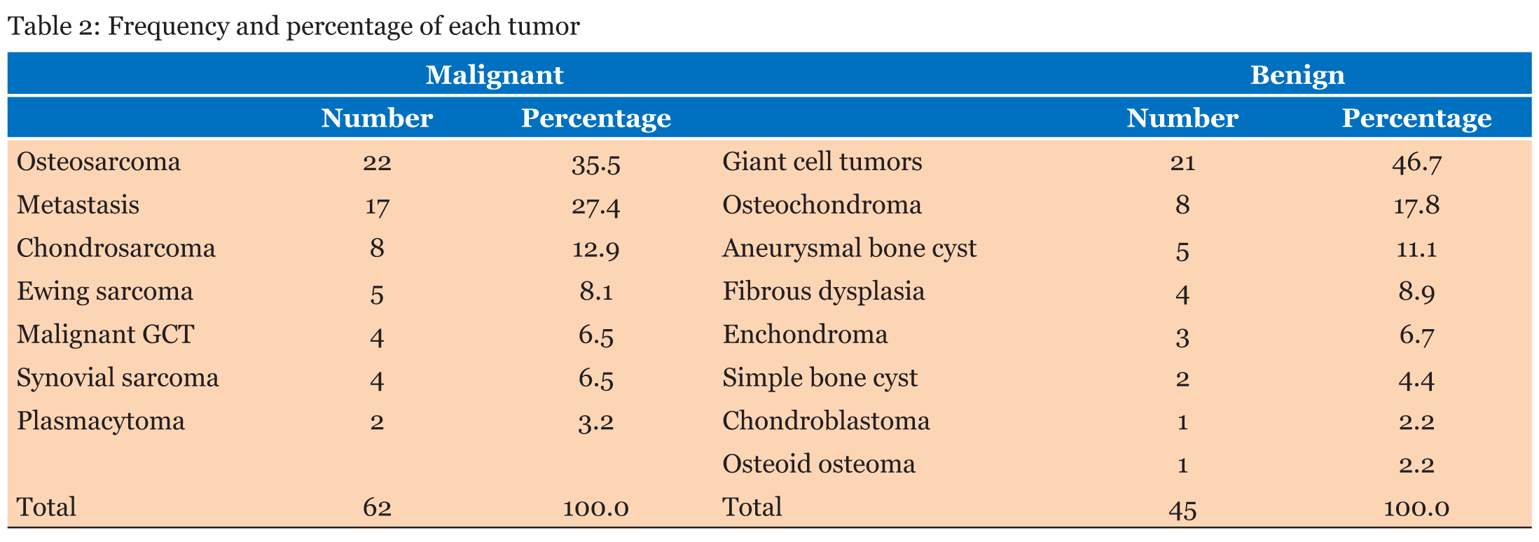
Results: Of all the 122 cases, 51.6% were affected in males while the rest 48.4% were affected in females. The first most common malignant bone tumors were osteosarcoma (22; 35.5%), while the first most common benign bone tumors were giant cell tumor (21; 48.9%). In malignant bone tumors followed by metastasis (17; 27.4%) and chondrosarcoma (7; 11.3%). In benign bone tumors followed by osteochondroma (8; 17.8%) and aneurysmal bone cyst (3; 6.7%). Femur was the most common site of malignant musculoskeletal tumors, followed by the humerus, while tibia is a common site for benign tumors.
Conclusion: Epidemiology of musculoskeletal tumors in Yogyakarta, in comparison with other parts of the world, are no significant differences. This report represents a first of its kind in our region, and gives representative results to be compared to other South East Asia, region.
Keywords: Benign, Epidemiology, Malignant, Musculoskeletal, Tumor
INTRODUCTION
One of the main causes of mortality in the world are malignant neoplasms. These neoplasms can occur in musculoskeletal system either as a primer or metastasis from other organs such as breast, kidney, lung and thyroid [1],[2],[3]. Diagnosis of neoplasms is very important because it determines subsequent management planning.
Primary bone tumors are often diagnosed late, due to their rare incidence, about 0.2–0.5% of all malignancies at all ages [4], with 3–5% of these tumors diagnosed in children under 15 and 7–8% between the ages of 15 and 19 years in European [5]. Signs and symptoms are often nonspecific. Usually suspected and treated as osteomyelitis or simple fracture before finally getting the right diagnosis. The incidence of primary bone tumors is about 9 in 1 million people worldwide, where in men slightly more common (10 in 1 million) than in women (8 in 1 million) [6]. To improve diagnosis and preliminary procedures, epidemiological studies are needed in each region. These studies will also help and guide the researchers to look for any specific risk factors in the area. Previous studies show Europe and USA have higher incidence and prevalence rates of primary bone tumor than Asia [5],[7],[8].
MATERIALS AND METHODS
We collected data on patients underwent either open or excisional musculoskeletal biopsies at Sardjito general hospital in Yogyakarta from January 2011 to December 2014, retrospectively. Data were then divided by sex, age, anatomical site and histopathologic type, whereby the diagnosis was upheld according to the results of clinicopathologic conferences according to clinical presentation and radiographic findings. Tumors are then classified into benign bone tumors, malignant bone tumors, and soft tissue tumors. Soft tissue tumors are just malignant ones such as fibrosarcoma, rhabdomyosarcoma, and so on. Whereas benign soft tissue tumors were not included.
RESULTS
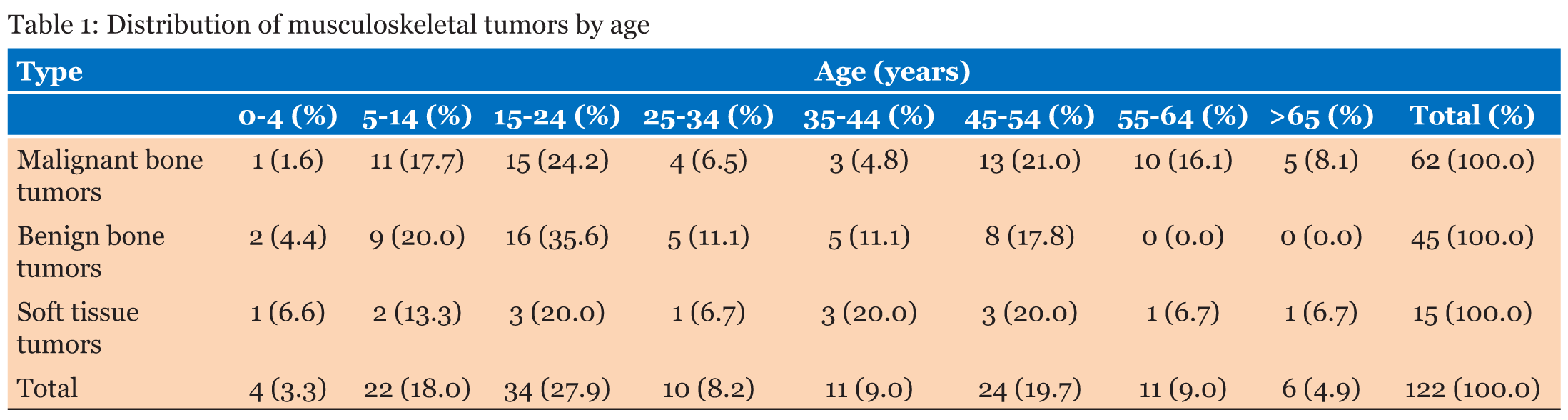
Totally, 122 cases were analyzed. Male to female ratio was 1.07. There were 59 women (48.4%) and 63 men (51.6%). The oldest patient was an 81-year-old man with metastasis. Table 1 shows that the most common anatomical site of bone tumors was the femur (38; 35.5%), followed by the tibia (21; 19.6%) and the humerus (17; 15.9%). The peak frequency in age of patients are between 5 and 25 years. And in patients younger than 25 years old shows a large population of benign bone tumors (27; 60.0%). In the age group of 35-54 years, soft tissue tumors occured more (6; 40.0%). The youngest patient reported was a 1-year-old child with chondroblastoma.
Benign bone tumors
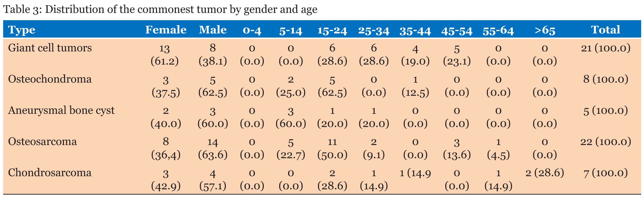
The giant cell tumor (22; 48.9%), osteochondroma (8; 17.8%), aneurysmal bone cyst (3; 6.7%) were the most frequently occuring tumors. In adults patients with third and fourth decades of life, giant cell tumors were seen mostly (Table 2). Table 3 shows osteochondroma and aneurysmal bone cyst are often affect men more as compared to women. While women are in higher risk (61.9%) of suffering from giant cell tumor. Also, about half of the cases of osteochondroma belonged to 15–25 years (62.5%) and aneurysmal bone cyst belonged to 5–14 years (60.0%).
Malignant bone tumors
The most common malignant neoplasms was osteosarcoma (22; 35.5%), followed by metastasis (17; 27.4%), and chondrosarcoma (7; 11.3%), of a total 62 malignant neoplasms. Men were more frequently affected by osteosarcoma and chondrosarcoma than women and more frequent in the 14–24 years age group. The ratio for osteosarcoma comparison in men as compared to women was 1.75, while the ratio for chondrosarcoma was 1.3 (Table 3).
Soft tissue tumors
The anatomic location of soft tissue tumors most commonly found on wrist-hand (5; 33.3%), thigh (4; 26.7%), elbow joint (4; 26.7%) and leg (2; 13.3%). Of all cases, 15 cases of malignancy in soft tissue were found, and women were more frequently affected (10; 66.7%) than men.
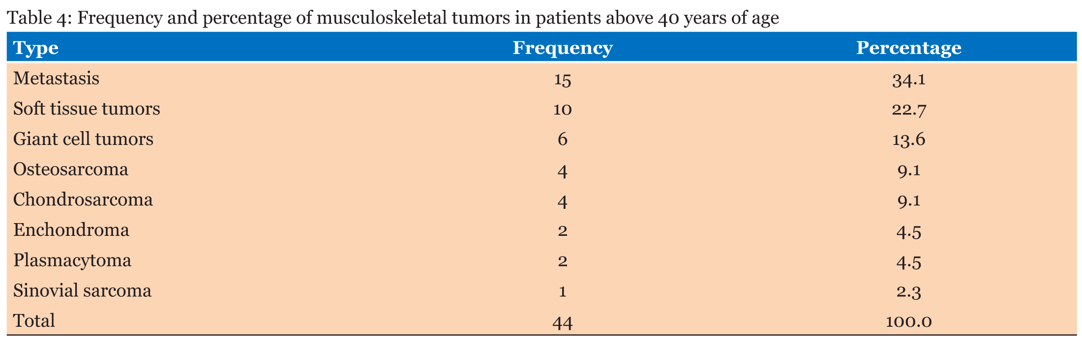
Musculoskeletal tumors in patients over 40 years
In this group, there were 44 patients, 23 (52.3%) were men and 21 (47.7%) were women.Table 4 shows that most malignat bone tumors are osteosarcoma (4; 9.1%), whereas benign bone tumors are mostly giant cell tumors (6; 13.6%). The table also shows the most pathology in this group is metastasis (15; 34.1%), followed by soft tissue tumors (10; 22.7%).
Anatomical site distribution
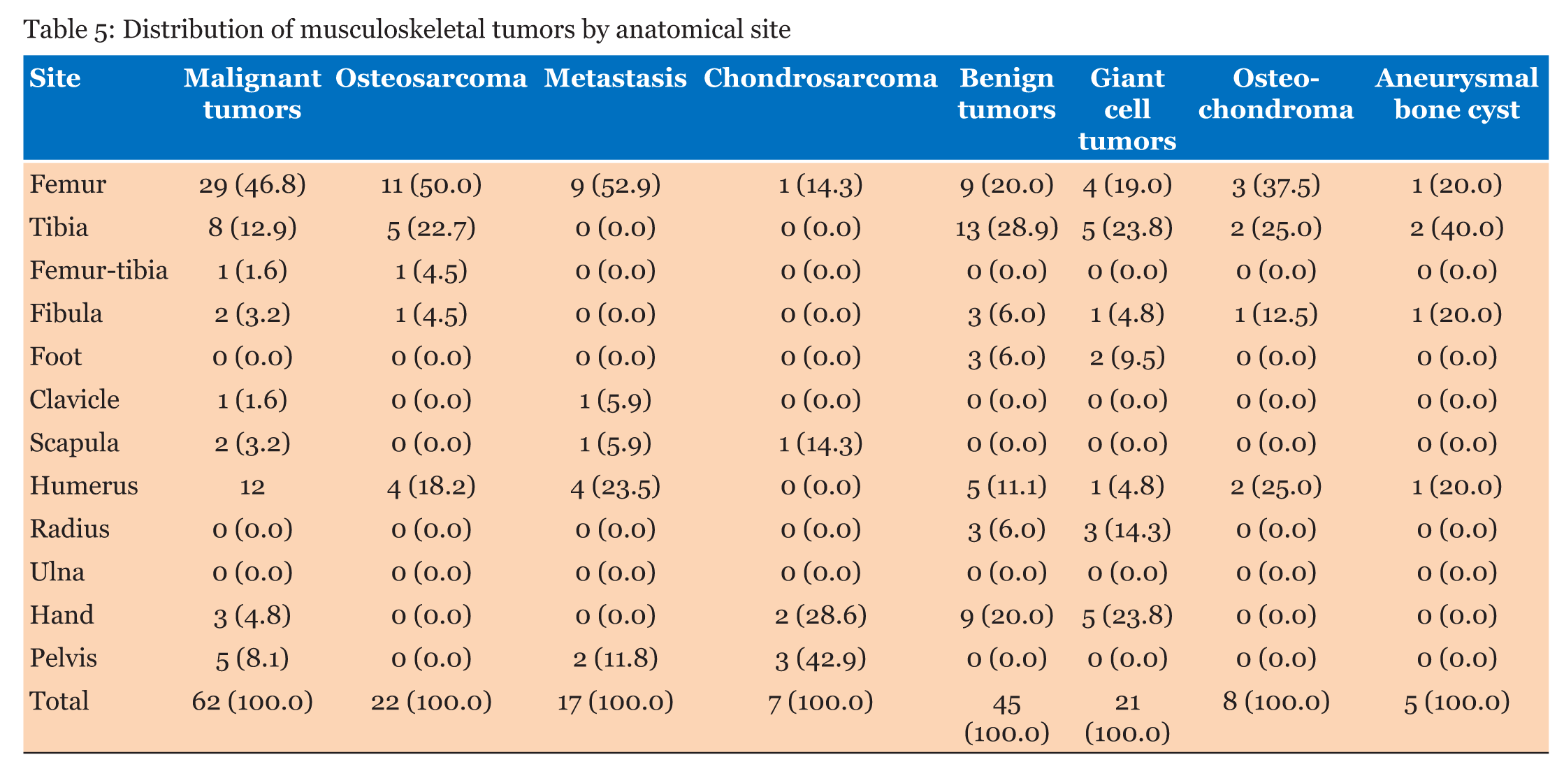
The most common site of malignant bone tumors in long bone involved was the femur, followed by the humerus (12; 19.4%) and the tibia (8; 12.9%). While in benign neoplasms, femur was also the most commont site involved, followed by the tibia (13; 28.9%) and the wrist-hand (9; 20.0%). Table 5 is illustrating anatomical site distribution of specific tumors, showing there is a case of osteosarcoma involving of more than one bone, the femur and the tibia.
DISCUSSION
Previous studies show that the relative 5-year survival rate is 53.9% for osteosarcoma, 75.2% for chondrosarcoma, and 50.6% for Ewing’s sarcoma [9]. Although primary bone tumors only contribute 1% of all malignancies [4],[10], they have a high mortality rate. Better diagnostic and management strategies require regional description and distribution data of musculoskeletal tumors that are one of the causes of morbidity and mortality worldwide. According to previous studies, osteosarcoma is still the most frequent primary bone malignancy [6]. It was reported that osteosarcoma contributed 35-68% of all malignant bone tumors [11],[12]. This is similar to what we found in our study in Yogyakarta, ososarcoma was found 35.5% of the total patients. The greatest number was found in age under 25 years (16; 72.7%) and there were no patients under five years of age. The ratio between men and women is the same as other studies which is about 1.75. Ranked second in the primary bone malignancy is chondrosarcoma (11.3%), followed by Ewing Sarcoma (4; 6.5%). Our results are similar to those reported in a study by Rao et al. [13] in India and by Shah et al. [11] in Pakistan. Most reports from developed countries explain chondrosarcoma as the second most frequent. Ewing’s sarcomas are tumors of the young, with 80% occurring in patients under 20 years. They rarely occur above the age of 30 [5]. We did not have any Ewing’s sarcoma patient older than 35 years. In our study, the most frequent benign bone tumor was giant cell tumor, followed by aneurysmal bone cyst and osteochondroma. Giant cell tumor accounted for 46.7% of benign bone tumors and 19.6% of all bone neoplasms. The second most frequent benign neoplasm, osteochondroma, accounted for about 17.8% of cases, wherein 62.5% cases were diagnosed in femur and tibia. Humerus was the next most common site. Giant cell tumors, as the second most common benign tumors in several studies [14],[15], represent about 5% of all bone tumors. In our study, it is the most frequent benign tumor. It was seen in 21 patients (19.6%) with involvement of the femur (19.0%), tibia (23.8%), and hand (23.8%) in order of frequency. The real frequency of this tumor may be more in our population because of its clinical diagnosis by radiographs.
Metastasis from other sites [2],[3] and multiple myeloma are the most likely diagnosed bone lesions in patients older than 40 [16]. Metastatic carcinoma and soft tissue tumors were the most prevalent diagnosed bone tumors in older patients. Giant cell tumor (6; 13.6%) and osteosarcoma (4; 9.1%) were the common primary benign and malignant bone tumors, respectively.
Late diagnosis and poor prognosis of musculoskeletal tumors are associated with some reasons. Nonspecific clinical symptoms, just like pain and swelling, which are more common in other musculoskeletal diseases than tumors, and unfamiliarity of general medical staff to the symptoms, besides the rarity of musculoskeletal tumors, are some of the causes. Epidemiologic studies based on e-databases in each region help in rapid diagnosis and management. This study has no significant difference related to epidemiology of musculoskeletal tumors with others in literature.
CONCLUSION
Epidemiology of musculoskeletal tumors in Yogyakarta, in comparison with other parts of the world, are no significant differences. This report represents a first of its kind in our region, and gives representative results to be compared to other South East Asia region.
REFERENCES
1.
Solooki S, Vosoughi AR, Masoomi V. Epidemiology of musculoskeletal tumors in Shiraz, South of Iran. Indian J Med Paediatr Oncol 2011 Oct;32(4):187–91. [CrossRef]
[Pubmed]

2.
Coleman RE. Clinical features of metastatic bone disease and risk of skeletal morbidity. Clin Cancer Res 2006 Oct 15;12(20 Pt 2):6243s–9s. [CrossRef]
[Pubmed]
3.
Lewis VO. What's new in musculoskeletal oncology. J Bone Joint Surg Am 2007 Jun;89(6):1399–407. [CrossRef]
[Pubmed]
4.
Fletcher CD, Unni KK, Mertens F. Pathology and Genetics of tumours of Soft tissue and Bone: World Health Organization Classification of tumours. Lyon, France: IARC Press, 2002.
5.
Stiller CA, Bielack SS, Jundt G, Steliarova-Foucher E. Bone tumours in European children and adolescents, 1978–1997. Report from the automated childhood cancer information system project. Eur J Cancer 2006 Sep;42(13):2124–35. [CrossRef]
[Pubmed]
6.
Bramer JAM, Somford MP. The epidemiology of primary skeletal malignancy. Orthopaedics and Trauma 2010;24(4):247–51. [CrossRef]
7.
Parkin DM, Stiller CA, Nectoux J. International variations in the incidence of childhood bone tumours. Int J Cancer 1993 Feb 1;53(3):371–6. [CrossRef]
[Pubmed]
8.
Eyre R, Feltbower RG, Mubwandarikwa E, Eden TO, McNally RJ. Epidemiology of bone tumours in children and young adults. Pediatr Blood Cancer 2009 Dec;53 (6):941–52. [CrossRef]
[Pubmed]
9.
Shah SH, Muzaffar S, Soomro IN, Pervez S, Hasan SH. Clinico-morphological pattern and frequency of bone cancer. J Pak Med Assoc 1999 May;49(5):110–2.
[Pubmed]
10.
Omololu AB, Ogunbiyi JO, Ogunlade SO, Alonge TO, Adebisi A, Akang EE. Primary malignant bone tumour in a tropical African University teaching hospital. West Afr J Med 2002 Oct–Dec;21(4):291–3.
[Pubmed]
11.
Rao VS, Pai MR, Rao RC, Adhikary MM. Incidence of primary bone tumours and tumour like lesions in and around Dakshina Kannada district of Karnataka. J Indian Med Assoc 1996 Mar;94(3):103–4, 121.
[Pubmed]
12.
Yeole BB, Jussawalla DJ. Descriptive epidemiology of bone cancer in greater Bombay. Indian J Cancer 1998 Sep;35(3):101–6.
[Pubmed]
13.
Damron TA, Ward WG, Stewart A. Osteosarcoma, chondrosarcoma, and Ewing's sarcoma: National cancer data base report. Clin Orthop Relat Res 2007 Jun;459:40–7. [CrossRef]
[Pubmed]
14.
Baena-Ocampo Ldel C, Ramirez-Perez E, Linares-Gonzalez LM, Delgado-Chavez R. Epidemiology of bone tumors in Mexico City: Retrospective clinicopathologic study of 566 patients at a referral institution. Ann Diagn Pathol 2009 Feb;13(1):16–21. [CrossRef]
[Pubmed]
15.
Odetayo OO. Pattern of bone tumours at the National Orthopaedic Hospital, Lagos, Nigeria. West Afr J Med 2001 Apr-Jun;20(2):161–4.
[Pubmed]
16.
Heck RK. General principles of tumors. In: canale St, Beaty JH, editors. campbell's Operative Orthopaedics. 11ed. Philadelphia: Mosby Elsevier; 2008. p. 775–854.
SUPPORTING INFORMATION
Author Contributions
Rahadyan Magetsari - Substantial contributions to conception and design, Acquisition of data, Analysis of data, Interpretation of data, Drafting the article, Revising it critically for important intellectual content, Final approval of the version to be published
Doni Agustian - Substantial contributions to conception and design, Acquisition of data, Analysis of data, Interpretation of data, Drafting the article, Revising it critically for important intellectual content, Final approval of the version to be published
Guaranter of SubmissionThe corresponding author is the guarantor of submission.
Source of SupportNone
Consent StatementWritten informed consent was obtained from the patient for publication of this study.
Data AvailabilityAll relevant data are within the paper and its Supporting Information files.
Conflict of InterestAuthors declare no conflict of interest.
Copyright© 2018 Rahadyan Magetsari et al. This article is distributed under the terms of Creative Commons Attribution License which permits unrestricted use, distribution and reproduction in any medium provided the original author(s) and original publisher are properly credited. Please see the copyright policy on the journal website for more information.
